The shape of the bottom part of the knee joint is emerging as an important predictor of whether someone who ruptures their ACL will repeat the injury after reconstruction and rehabilitation.
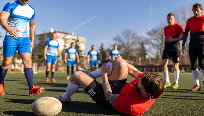
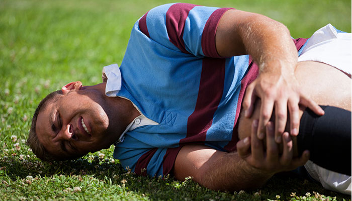
The ACL is one of the main ligaments stabilising the knee joint. Ruptures, or tears, are common injuries in all codes of football, as well as netball and basketball, and they usually happen when a player lands badly from a jump, twists, or changes direction quickly.
A ruptured ACL can be surgically reconstructed by replacing the damaged ligament with a tendon taken from elsewhere in the patient’s body, such as a hamstring, but they cannot return to sport for 12 months.
One severe ACL injury can be career-ending, but up to 35 per cent of all ACL reconstructions fail, with patients potentially going on to suffer a second or even a third injury.
As a former rugby player, specialist orthopaedic surgeon Dr Michael Dan has seen many careers ended by knee injuries.
In dogs, the cranial cruciate ligament (CCL) performs a similar function to the ACL in humans, and like humans, injuries are common and often require surgical intervention.
Before beginning his medical training, Dr Dan played for Australia in the Under 21s World Cup, going on to be signed to the Western Force rugby team in Western Australia.
“With every ACL reconstruction, the likelihood the player will return to their sport drops,” Dr Dan says.
“That’s devastating at any stage of your career, but these repeat injuries are particularly common in young men in their late teens and early 20s – just when they’re just coming into their peak.
“When you combine high rates of participation in pivoting sports with the sense of being bulletproof that we can have at that age, the increased rate of reinjury isn’t really that surprising.
“Female participation and exposure to professional sporting environments is increasing, and young women now seem to be overtaking their male counterparts with regards the incidence of ACL injuries.”
Tibial plateau angle as a predictor
The tibial plateau is the top surface of the tibia – the heavier of the two bones in the lower leg. This is where the femur rests, forming the knee joint.
Observed from the side, the tibial plateau has a visible slope, and in most people, the angle between the two bones is between 6 and 12 degrees. In some people, however, the tibial slope is steeper, and each extra degree means more pressure is being exerted on the ACL.
“We’re beginning to collect data now on these repeat cases, and so far, that is pointing to reinjury being most likely in patients with a higher tibial plateau angle,” Dr Dan says.
“In dogs, the cranial cruciate ligament (CCL) performs a similar function to the ACL in humans, and like humans, injuries are common and often required surgical intervention.
“To reduce the likelihood of the failure of CCL reconstructions, veterinary surgeons reduce the angle at which the bones meet by perform an osteotomy, which involves removing a small wedge of bone.
“This puts less pressure on the ligament, making a second rupture less likely without affecting the operation of the joint.”
For dogs, this has been the main treatment since the early 1990s.
Bringing a French gold standard to Australia
French orthopaedic surgeons pioneered a similar procedure for human patients in 1991 for people who had had multiple ACL failures.
It has now evolved to become more common, and is now performed on an almost weekly basis in France. In some cases, it is performed at the same time as the first reconstruction.
Dr Dan was first exposed to the slope-levelling osteotomy in its earlier days, while a friend was playing for a French rugby team.
One of his friends, a champion player who was contracted to a club in Lyon, suffered a third ACL injury that looked certain to sideline him permanently.
The club encouraged him to see their own surgeon, who reconstructed the ACL and performed an osteotomy. He made a complete recovery and went on to play for another seven years before retiring in his early 30s.
Completing his own orthopaedic training, Dr Dan discovered the procedure was uncommon in Australia, so he returned to France to complete fellowship training in complex knee surgery at the Lyon Knee School.
He is continuing to refine the process he learned, now using low-radiation EOS scanning to accurately measure the angle of a patient’s tibial slope and exactly how much bone needs to be removed and from where.
“I think a lot of Australian orthopaedic surgeons either aren’t familiar with tibial slope osteotomy and its benefits, or they think of it as too risky because they haven’t observed its development and natural progression over the past 30 years,” he says.
“It’s something I believe we should be doing more of in Australia, particularly when someone has a higher-than-average tibial plateau angle and is potentially more vulnerable to reinjury.
“It’s hugely beneficial, particularly for teenagers who have a high likelihood of re-rupture and their whole sporting careers ahead of them.
“Why should we put these individuals with a high tibial slope through a standard ACL reconstruction on its own when we know it has such a low chance of success?
“If we can do it once and do it right by putting the new ligament in the optimal biomechanical environment, that's going to be advantageous.”
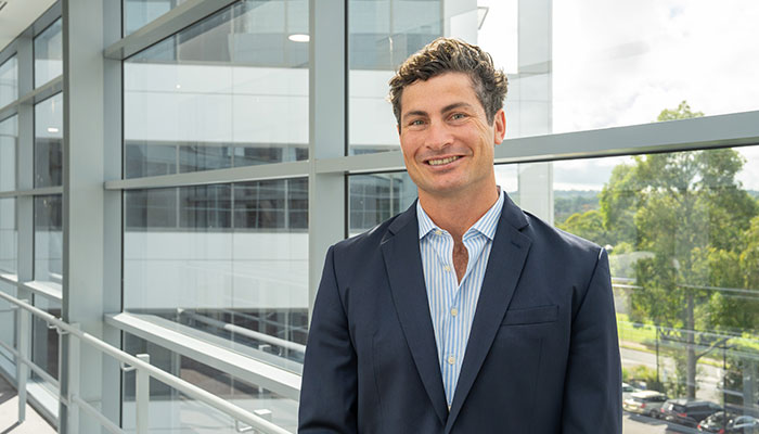
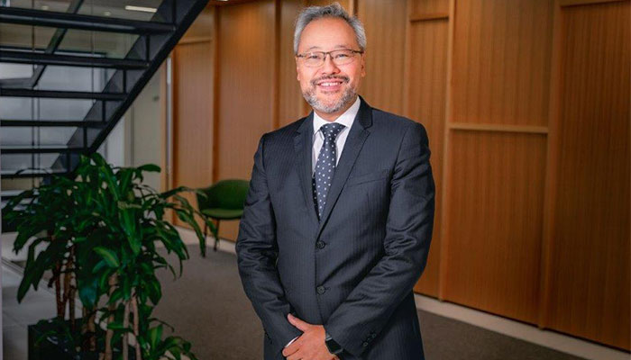
Dr Michael Dan, pictured above, is an orthopaedic surgeon specialising in complex lower limb ligament and tendon problems. In Sydney, he practices at Macquarie University Hospital and Clinics.

