In a new paper published in international journal, Nucleic Acids Research, a team of researchers from the Motor Neuron Disease Research Centre at Macquarie University described TDP-43 changing its structure and forming specialised droplets in the spinal neurons of living zebrafish for the first time.
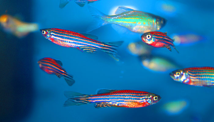
Crucial: Zebrafish have been instrumental in neurodegenerative disease research for many years because they have a very similar genome to humans.
TDP-43 is a protein produced in the nucleus of cells in the human brain and central nervous system, including the motor neurons that control movement.
Under normal conditions, it is vital to the healthy functioning of these cells, but when it becomes pathogenic, it leaves the nucleus and begins to form solid clumps in the surrounding cell body, or cytoplasm, clogging it and preventing the cell from functioning properly.
These solid clumps are present in 97 per cent of cases of MND (with amyotrophic lateral sclerosis or ALS being the most prominent form of MND), and in some types of dementia.
Breaking them down, and preventing them from forming in the first place, is the subject of research worldwide.
An easy way to think of it is as a sort of vinaigrette: shake the vinaigrette and the oil mixes with the vinegar to form an emulsion, but stop shaking and the oil settles back into droplets after a while.
The paper’s senior author, Associate Professor Marco Morsch, says the team harnessed a process known as phase separation to observe the way the protein shifted between different states, and moved from the cell nucleus into the cytoplasm.
“TDP-43 is a dynamic protein that is dispersed throughout the nucleus when it is doing its work, but forms into droplets when it is not,” he says.
“An easy way to think of it is as a sort of vinaigrette: shake the vinaigrette and the oil mixes with the vinegar to form an emulsion, but stop shaking and the oil settles back into droplets after a while.
“Normal TDP-43 shifts back and forth between droplet and liquid, but pathogenic TDP-43 droplets may not shift back the same way. For example, when they lose their ability to bind to RNA in the nucleus, they may be more prone to form hardened clumps in the cytoplasm.”
The team is now seeking funding to use this information to develop ways of keeping TDP-43 in its liquid-like state.
Once they understand these principles better, they hope to look at modifying certain regions of the protein, or even developing anti-clumping instructions.
Zebrafish and their importance in neurodegenerative disease research
The Macquarie researchers introduced normal and pathogenic human proteins into the spinal cords of zebrafish in order to observe the proteins’ behaviour.
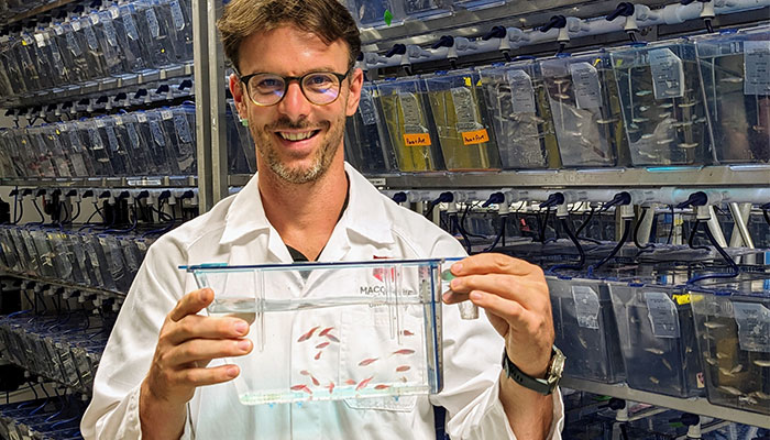
Hope: Associate Professor Marco Morsch, pictured above, says their breakthrough could speed the development of treatments for MND and dementia.
Previous studies have been able to recreate TDP-43 clumping in vitro, but this is the first time that this dynamic process has been characterised in such detail in a living vertebrate.
Also known as danios, zebrafish are a popular choice for aquarium owners. They have been instrumental in neurodegenerative disease research for many years because they have a very similar genome to humans, sharing 86 per cent or more of our disease-causing genes.
We also share similar cell machinery and basic biological components. In humans, motor neurons control the muscles in our arms and legs, while in zebrafish, they drive the constant movement of their fins.
As adults, zebrafish can grow up to four or five centimetres, but three days after hatching, they are just a few millimetres long, and breathe by absorbing oxygen molecules from the water through their skin.
Importantly, the tiny hatchlings are also transparent, giving neuroscientists a literal window into an operating central nervous system that is extremely difficult to achieve with other vertebrates.
The team was able to observe the juvenile zebrafish under the microscope using a gel that kept them still while allowing them to breathe.
- How three hours could add up to a better retirement
- Review: What if Juliet didn't die at the end of Romeo and Juliet?
Associate Professor Morsch says their discoveries will be important for researchers worldwide, as it will improve testing of new drugs.
“There is an enormous amount of work underway on developing potential treatments that could prevent TDP-43 from clumping, or break down aggregates that have already formed,” he says.
“We have developed a platform that researchers can use to test these treatments by introducing them to the water, so the fish absorb them through their skin.
“It will be possible to see in real time whether something is working, which will make pre-clinical testing quicker and easier.
“We hope this could speed the development of treatments for diseases like MND and dementia, and help bring them to human trials more quickly.”
Associate Professor Marco Morsch is a neuroscientist and Co-Director of Research in the Motor Neuron Disease Research Centre at Macquarie University.
This work was supported by the ALS Foundation Netherlands, FightMND, and donations towards MND research at Macquarie University and the other research teams.
Research reported in this publication was supported by the National Institute on Drug Abuse, the National Institute of Mental Health (NIMH), the National Institute of Neurological Disorders and Stroke (NINDS) of the National Institutes of Health.