Macquarie Medical School Professor of Radiology, John Magnussen, spent four years working on The Mummy Project as part of a multidisciplinary team that included British Museum curator Dr James Fraser and Egyptologist Dr Conni Lord from the Chau Chak Wing Museum at the University of Sydney.
The team’s fascinating findings are documented in the new book, Speak My Name: Investigating Egyptian Mummies, which takes its title from a plea commonly written on the walls of Egyptian tombs: “Speak my name that I may live again”.
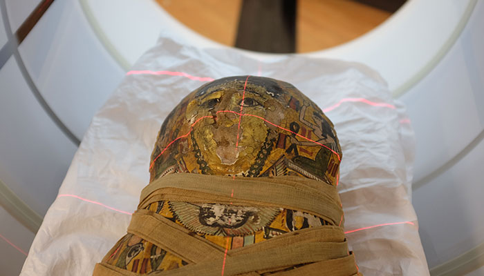
As part of the project, Professor Magnussen produced computed tomography (CT) scans of the four mummies and three coffins from the Chau Chak Wing Museum, revealing minute details that had never been seen before and allowing for a recreation of the weathered paintwork of one of the coffins.
Bringing modern science to ancient history
In the mid-19th century, Europe was in the grip of an obsession with ancient Egypt, and it was common for travellers to buy mummies and other artefacts.
Often, the mummies would be unwrapped, either for study or entertainment.
“Unwrapping a mummy is a one-way trip, rather like breaking an egg,” Magnussen says. “You might try to put it back together afterwards, but it will never be the same.”
With modern imaging equipment, a mummy can be scanned in the same way as a living body, showing it in great detail without damaging it.
CT scans and x-rays are the preferred methods, as magnetic resonance imaging (MRI) has the potential to show greater detail in scanned objects, but it is reliant on the presence of water particles. Living bodies are up to 70 per cent water, but mummies are very dry.
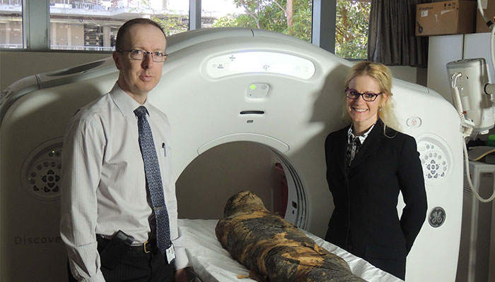
Wonderful adventure: Professors John Magnussen and Ronika Power, pictured, with the Macquarie Medical Imaging CT scanner used to reveal new details about ancient mummies housed in Sydney's Chau Chak Wing Museum.
In his career as a medical radiologist, Magnussen specialises in neuroradiology and cardiac imaging, but for the past 15 years, he has also been lending his expertise to the examination of ancient artefacts.
“Working with mummies is a wonderful adventure,” he says.
“The first time I had the opportunity to scan one, I was hooked. After that, people heard that I had an interest in the area and came looking for me.
“I think Egypt has been an ongoing source of fascination in modern times because it’s such a heady combination of mystery, history and privilege.
“Peering into a burial chamber dating back thousands of years is like touching greatness and experiencing fame.
“Most people’s bodies would simply have been wrapped and buried in the desert to dry out.
“The mummies that have survived were those of the elite. This level of care and artistry in death was very expensive – as much as thousands of ordinary Egyptians would have earned in their entire lives.
“They would have been the celebrities of their time.”
What we’ve done here is produce a digital archive so we can continue to remember and learn from these people. In a sense, it lets them to ‘live’ on, thousands of years after their deaths.
Since his first brush with ancient history, Magnussen has produced images of a number of human mummies, Babylonian clay tablets, Roman amphorae, and even a mummified cat.
“Mummified animals were frequently placed in tombs in Egypt, and cats were in great demand as they were considered sacred,” he says.
“When we scanned the cat mummy, we found it didn’t contain very much cat at all – it was mostly pieces of wood and stuffing, beautifully shaped to resemble a cat. Cats were in short supply, so dealers got creative.
“Mummies are a perfect example of not judging a book by its cover: the inside often bears little resemblance to the outside, and that’s part of the adventure.”
Looking beneath the surface
The four mummies and three coffins examined as part of The Mummy Project were purchased in 1856-7 by Sir Charles Nicholson, and now form part of the largest collection of ancient Egyptian artefacts in Australia.
Revelations: applying modern science to ancient artefacts has uncovered minute details that have never been seen before.
Under investigation were a coffin belonging to a woman named Meruah and the female mummy inside it dating about 200 years earlier, the coffin of a priest named Padiashaikhet and a Roman-period female mummy inside, the coffin of a woman named Mer-Neith-it-es and her jumbled remains inside, and the mummy of a young boy named Horus, who died about 100 CE when coffins were no longer commonly used.
A mismatch between coffin and mummy is not unusual, as antiquities dealers would often pair an empty coffin with an unrelated mummy to get a better price.
One of the main aims of the project was to determine the condition of the remains in Mer-Neith-it-es’s 2,500-year-old coffin. For decades, it had been believed to be empty, but it proved to contain the shattered remnants of a mummy that had been literally torn apart by grave robbers.
As damaged as the remains were, they presented a rare opportunity; most ancient coffins not holding complete mummies have had their contents discarded.
Scanning found the remains were so badly damaged that removing them could not damage them further. Painstaking excavations found the contents included a pair of mummified feet and more than 7,000 tiny faience beads that had probably been woven into a shroud.
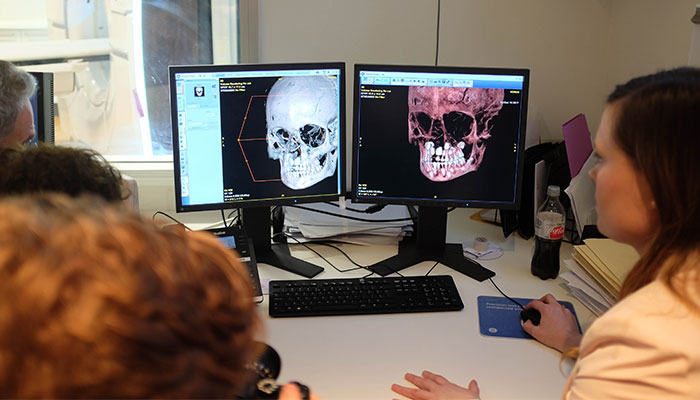
Poignant discovery: A mummy of the young boy, Horus, who is thought to have been aged seven or eight when he died in 100 CE.
Investigations to date have not been able to reveal a definitive cause of death for any of the mummies though the latest scans showed the mummy in Padiashaikhet’s coffin had her hands and toes sliced off to fit into the narrow box.
Magnussen says he found the images of the young boy the most poignant as his own children were of a similar age at the time.
“He was between seven and eight years old when he died, and looking at his skull, you can clearly see the adult teeth erupting from his jaw,” Magnussen says.
- Zebra birds are social singers: new study
- What linguistics can teach us about how to talk to people with dementia
“That had an impact on everyone in the room. It was something we were all familiar with.
“It may be odd, but I think these images are especially beautiful.
“What we’ve done here is produce a digital archive so we can continue to remember and learn from these people. In a sense, it lets them to ‘live’ on, thousands of years after their deaths.”
John Magnussen is a Professor of Radiology at Macquarie Medical School in the Faculty of Medicine, Health and Human Sciences.
Speak My Name: Investigating Egyptian Mummies is edited by James Fraser, Conni Lord and John Magnussen and published by Sydney University Press
A number of Macquarie University researchers, students and graduates were involved in The Mummy Project and contributed to Speak My Name: Professor Ronika Power, Professor Paul Haynes, Associate Professor Boyo Ockinga, Dr Karin Sowada, Dr Alice McClymont, Dr Michelle Whitford, Dylan Multari, Prathiba Ravishankar and Kate Gosford.
Macquarie Medical Imaging (MMI), where the Chau Chak Wing mummies were scanned, is a Node of the National Imaging Facility. Its state-of-the-art equipment is available for traditional diagnostic purposes as well as a variety of medical and scientific research.
Buy the Book here: https://sydneyuniversitypress.com.au/products/13106b